Chapter 4 : Motor Control

Brain: Contents Page


Vestibular Pathways and Balance

The Sense of Balance

Objects fall over if the centre of gravity is outside the base of the object; (the centre of gravity is the point through which the force of gravity acts). In the body, adjustments of the distribution of forces generated by muscles (‘Muscle Tone’) keep the line of gravity within the base.

Postural Responses : One way of dealing with forces that move the centre of gravity outside the base is to broaden the base - by separating the feet; or by side-stepping. Upright Posture and Balance are maintained by automatic adjustments of muscle tone; such movements are involuntary (they are not willed) and determine one’s posture.

Reflexes influencing balance. The sensory input that influences these adjustments comes from several sources:

  • the inner ear (utricle and saccule) senses the direction of gravity - the position of the head in space
  • the inner ear (semicircular canals) senses the direction of rotation or acceleration of the head, and provides some rapid dynamic responses
  • muscle, tendon and joint receptors sense joint the position of the limb
  • the eyes sense the visual environment, e.g. the horizon, and the distance of objects

All of these influence the distribution of forces generated in the body musculature, by sending information to the brainstem and cerebellum through which precise adjustments of muscle tone within the body are performed. 

Top

Equilibrium and the Responses to Tilting the Body

Sense of Equilibrium

The inner ear contains several sensory organs two of which provide information about the direction of the force of gravity. These are the Saccule and Utricle, and have been considered in the main site

These organs sense the direction of tilting of the head and produce some basic reflex (automatic) responses.

This reflex response involves the descending pathways from the vestibular nuclei, and these projections to the spinal cord are responsible for adjusting the position and tone of the muscles of the limbs and the axial muscles so as to keep the centre of gravity within the base.

If the tilt is sideways, the reflex produces some side-stepping in order to a wide base beneath the centre of gravity.

*


Tilting and Eye Movements

When the head is tilted, the eyes also rotate in the opposite direction, so as to maintain a horizonal horizon in the visual fields

This reflex response involves a short chain of neurones, starting in the vestibular apparatus and vestibular nuclei.

On each side of the midline of the brainstem, a band of axons called the medial longitudinal bundle and communicates information concerned with the position the head to the nuclei of the oculomotor, trochlear and abducent nerves, and to the paramedian pontine reticular formation (PPRF).

These pathways are responsible for the vestibulo-ocular reflexes, and the PPRF is concerned with coordinating eye movements in response to vestibular and other inputs.


*

intranet.tdmu.edu.ua

Top


Vestibular influences on the body musculature.

Vestibular Apparatus and the Control of Muscle Tone

Depending on the inputs from the vestibular apparatus, compensatory changes in muscle tone throughout the body are transmitted from the vestibular nuclei of the brainstem to the motoneurones in spinal cord using the vestibulospinal tracts. The neurones involved have cell bodies in the vestibular nuclei in the medulla, and long axons that conducts action potentials to the interneurones and motoneurones in the spinal cord.

Vestibulospinal tracts.

The vestibular nuclei give rise to the medial and lateral vestibulospinal tracts. The medial vestibulospinal tract descends in the anterior funiculus and innervates the upper half of the cord (above T6). It innervates neck muscles and stabilises the head on with respect to the trunk during changes of position of the body. It works in partnership with pathways that control the position of the head and eye movements (see above).

The lateral vestibulospinal tract projects ipsilaterally through the anterior funiculus of the spinal cord to all segments of the spinal cord. It increases the tone of antigravity muscles and is concerned with compensatory adjustments in posture to accommodate tilts and movements of the body.

*

The vestibulospinal tracts help to maintain balance, as a result of inputs from the vestibular apparatus that signal changes in the position or the rotation of the head. The vestibulospinal pathways adjust the tone of the musculature to compensate for movements that would otherwise affect balance, and this is done by modifying the excitation of different motoneurone groups in the spinal cord.

Pixshark.com

Top


Physiological Responses to Rotation.

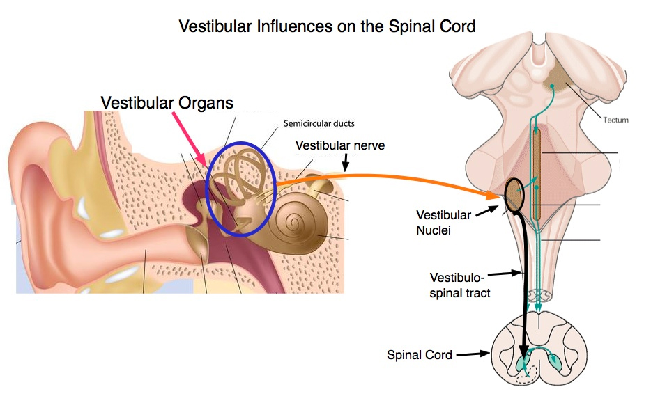
The detection of sudden movements (accelerations) to the head is performed by the three Semi-Circular Canals. This information is sent to the brainstem, to and induces sudden changes in posture that compensate for the acceleration.

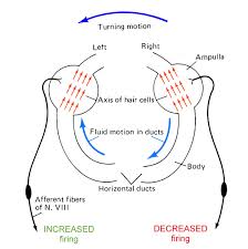
The diagram opposite shows that when the head is rotated in one direction the semicirular canals on both sides experience movement of the endolymph in the opposite derection. One set of hair cells bends so as to increase the dischearge in the vestibular nerve on that side. But on the opposite side, the tonic discharge arising from that ampulla is inhibited.

*

Nystagmus - 'Dancing Eyes'- is an involuntary eye movement consisting of a rapid movement of the eyes in one direction, followed by a slow movement back to the starting position.

Physiological Nystagmus

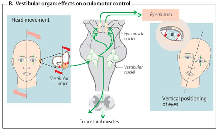
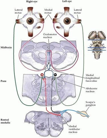
When the head is rotated, distant visual images would move beyond the visual field very rapidly, and reflexes initiated by the semicircular canals attempt to fixate the gaze on the moving object. This involves a slow movement of the eyes in the direction opposite to the rotation, during which the retina fixates and follows the distant object, followed by a rapid movement of the eyes in the direction of rotation after whch the eye fixates on a new distant object, and follows it.

The semicircular canals sense angular momentum, and send information to the vestibular nuclei. The medial longitudinal fasiculus (or bundle) runs up the brainstem near the midline below the fourth ventricle and makes contact with the cranial nerve nuclei that cause the extraocular muscles peroform the tasks mentioned above. The direction of ocular movement depends on which semicircular canals are activated by the rotation.

Pathological Nystagmus

Pathological nystagmus may be caused by congenital disorders, acquired or central nervous system disorders and drugs such as alcohol. The CNS disorders include disorders of the vestibular system, brainstem or cerebellum (particularly the flooculo-nodular lobe). Nystagmus is sometimes associated with vertigo, a sense of dizziness accompanied by a sensation of rotatory movement.

The vestibulo-ocular reflexes can be tested using caloric testing, in which warm or cold water is passed into the external auditory canal. The change in temperature initiates convection currents in an adjacent semicircular canal, causes movements of the crista ampullaris, and initiates activity in the medial longitudinal bundle, producing nystagmus.

Eye Movements

The Frontal Eye Fields are an area (Brodmann's area 8) of each frontal lobe, which whe stimulated electrically result in coordinated movements of both eyes in a horizontal direction.

This area receives input from the visual asssociation cortex and connects with the superior colliculi and the paramedian pontine reticular formation (PPRF), a region of the midbrain that has vestibular inputs and is involved in coordinating eye movements.

The superior colliculi are involved in initiating changes in the diretion of gaze when objects suddenly move into the periphery of the visual fields.

Vestibular Connections with the Cerebellum

The cerebellum is concerned with the control of movement, integrating signals from different parts of the nervous system and generating error signals that allow adjustments to be made to any muscles involved so as to achieve the desired objective of the movement.

The vestibular apparatus and the vestibular nuclei have a special relationship with the oldest part of the cerebellum, the flocculonodular lobe. This lobe modulates the vestibulo-ocular reflex system and is used to help stabilize the position of the eyes during rotation and tilting. Neurons in this part of the cerebellum extract from the vestibular input a signal about the speed of movement that allows the eyes to follow a moving object smoothly - this is known as a pursuit movement of the eyes. Visual inputs to the cerebellum can modify these pursuit movements.

Some authors argue that the flocculonodular lobe plays an important role in the ability for the vestibular system to adapt to changes in the visual environment, and that this adaptation is important for the development of motor skills.

Motor memory can be attributed to pathways being laid down in this lobe of the cerebellum during the early development of motor skills. The ability of a child to sit up, stand, balance and walk appear to be dependent on pathways laid down in this part of the cerebellum during development.

These cerebellar circuits also allow the cerebellum to adapt to and compensate for damage to the inner ear. This compensation only occurs if the connections between the vestibular nerves and the flocculonoduar lobe are intact, and involve modulation of the vestibulo-ocular reflex pathways. Damage to this region will result in vertigo and/or nystagmus.

kin450-neurophysiology.wikispaces.com
www.d.umn.edu

Top


Control of the Intrinsic Eye Muscles.

The control of the intrinsic muscles of the eye is not concerned with Balance or Eye movements, but an important component of this innervation is carried by the oculomotor nerve, which is important in the control of extraocular muscles..

Control of the Intrinsic Muscles of the Eye

The Oculomotor Nerve (III) arises from the midbrain as shown opposite. The motoneurones, shown in red, innervate the extra-ocular muscles which are responsible for movements of the eyeball.

The autonomic (parasympathetic) fibres of the III nerve originate in the Edinger-Westphal nucleus and travel to the ciliary ganglia close to the eyeballs. There they synapse with post-ganglionic parasympathetic neurones that innervate the iris and the ciliary muscles of the anterior chamber of the eye.

The intrinsic muscle of the eye also have a sympathetic nerve supply consisting of the postganglionic neurones of the superior cervical ganglion.

*
Pixshark.com

Top

Chapter 4 : Motor Control

Brain: Contents Page


© HumanPhysiology.Academy 2014-2015